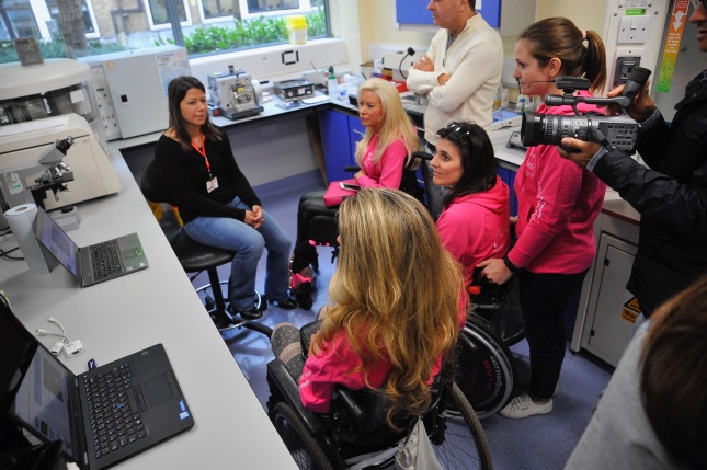
On January 20th 2014 the Cure Girls went to visit the labs of pharmacology of the university of Milan and met with Dr. Daniele Bottai. He showed us the new labs and gave us an interview to explain what his team is working on with regards to a cure for SCI.
1. Can you briefly describe the research that you are doing in relation to spinal cord injury?
When in 2006, I moved at the University of Milan, I have begun to get interested in Spinal Cord Injury (SCI) using a new (at the time) “drug”: neural stem cells. Between 2002 and 2006, I worked in the Laboratory of Professor Angelo Vescovi where I learned to manipulate neural stem cells (from different regions of the brain) both human and mouse.
In these 8 years spent to the University of Milan we have been studying the role of different types of stem cell in transplantation in animal model of SCI, in particular, we have studied the effects of murine embryonic and neural stem cells and human amniotic fluid (AFCS ) with the purpose to find the sources of stem cells that were an available source and with the appropriate characteristics for the treatment of neurological diseases .
In general, we can say that these cellular processes (performed in acute spinal cord lesion) have positive effects and are significant from a functional and morphological point of view. After treatment with the cells listed above, the mice returned to walk (albeit not the same as they did before the lesion); while lesioned not treated animals are able to move their hind limbs, but not to walk. (These results have been summarized in three scientific papers:
D. Bottai, D. Cigognini, L. Madaschi, R. Adami, E. Nicora, M. Menarini, A.M. Di Giulio, A. Gorio (2010).Embryonic Stem Cells Promote Motor Recovery and Affect Inflammatory Cell Infiltration in the Spinal Cord Injured Mice Experimental Neurology 223, 452-463 ;
D. Bottai , L. Madaschi, A.M. Di Giulio and A. Gorio. (2008) Viability -Dependent Promoting Action of Adult Neural Precursors in Spinal Cord Injury . Molecular Medicine , 14 (9-10), 634-644.
On the bases of these results, we asked what was the mechanism that caused this improvement.
The answer was that in this model the role of animal cells is purely trophic and they are not going to replace, if not in small portion, damaged or dead cells.
Various are the trophic molecules (cytokines) that are involved in this phenomenon. Recently, we have focused our attention on amniotic fluid cells. We chose this cell type because their availability since the at term cesarean delivery could represent an unlimited source of stem cells with no ethical issues and few risks for the child and the mother.
In a work that a few days ago has been accepted for publication (D. Bottai , G. Scesa , D. Cigognini, R. Adami , E.. Nicora, S. Abrignani, A.M. Di Giulio, and A. Gorio Third trimester amniotic fluid NG2 -positive cells are effective in improving on repair in spinal cord injury. Experimental Neurology.) we have shown that a trophic factor, produced by the AFCS, which is important for the induction of the morphofunctional recovery, was the hepatocyte growth factor (HGF) and that this cytokine was produced only by particular sub- populations of our cells or those expressing on their surface the NG2 protein (a membrane proteoglycan). Such a membrane protein can be hopefully used in the future to select from the amniotic fluid liquid the cells that express NG2 and so have a therapeutic action.
We are currently investigating what is the correlation between NG2 and HGF.
2. Acute injury or chronic injury present any difference for a research approach? Could you explains the differences and the advantages and disadvantages.
I do not think we can talk about differences between acute and chronic lesion in terms of advantages and disadvantages. These are two different pathological conditions, the acute progress into the chronic with the passage of time mostly because there is a de- myelination process.
In this context, we are dealing with two different types of patients the acute ones, namely that a few days or weeks have suffered damage to the spinal cord which have a very extensive inflammatory condition that exacerbates the primary mechanical injury further damaging the tissue and those, who instead, underwent chronic spinal cord injury for more time (months and years) in which degeneration induced by primary damage and the secondary SCI causes the formation of a cavity surrounded by the glial scar that separates the lesion from the undamaged tissue and prevent nerve regeneration.
Currently the researcher and clinician are faced with these types of patients because in the first instance they have not been able to prepare effective therapies to treat acute patients.
The therapeutic approach to the patient who recently underwent spinal damage is intended to reduce the compression state and to control the secondary damage due to inflammation through the drug methylprednisolone, inter alia, that approach does not seem to have a sufficient efficacy, as evidenced by the fact that the number of chronically para or quadriplegics is unfortunately increasing.
In the chronic patient instead we find ourselves facing a very different situation with the blood-brain barrier that is closed, and a glial scar consisting mainly of fibroblasts from the meninges and reactive astrocytes that produce proteoglycans (extracellular matrix molecules) that are responsible for the inhibition growth of axons.
In this situation, the therapeutic approach is vastly different from that prepared in the state of acute spinal cord injury.
In fact, removal (either mechanical – surgical or enzymatic) is a sine qua non con-diction in order to prepare any kind of intervention to restore or replace dead or damaged cells and rebuild the axons making them grow in the appropriate direction.
In this context, the treatment of chronic patients need multiple concurrent interventions:
- Treatment with drugs that induce axonal regeneration;
- Treatment with drugs that reduce the inhibitory effects of glial scar both mechanical and enzymatic chondroitinase that due to factors such as chemical inhibitors, blockers of Nogo and other myelin components;
- Treatment with cells or systems consisting of nanomaterials and cells.
Some of these approaches were ineffective few years ago but in the light of developments in nanomaterials and new types of stem cells I think it might be appropriate to re-examine these pathways.
3. How do you think we can solve the problem of scar?
As I mentioned in the previous answer, in order to find an approach that leads to the recovery of sensory and motor pathways, it is necessary to make the scar area permissive for the survival of cells that are transplanted and that would allow axonal growth in the manner to ensure the recovery of the routes of transmission.
With this in mind, scar removal is definitely needed and should be prepared to minimize the risk of inducing further damage.
In this context, the experimental work in the preclinical phase or with animal models is essential but at the same time very complex given that small animals have practical difficulties of intervention and larger animals have problems is housing costs that are beyond the economical capability of most the laboratories that I know.
Finally, the translation of the results obtained in the preclinical stage is very difficult for a variety of patients such as those with spinal cord injury whose disease is highly variable due to the fact that the damage is very random and therefore leads to differences between the patient and the other.
4. Can you apply your research to Chronic Spinal Cord Injury?
The applicability of the cells in the amniotic fluid in models of chronic spinal cord injury must be verified experimentally, so I can say a priori that such an intervention can be prepared but obviously need the appropriate adjustments of the experimental protocol. In fact, while in the acute model we have a purely trophic action in the case of chronic treatment the intervention should be at the level of local scar in order to determine whether these cells could contribute to modify the scar itself and reconstitute the ways by means of the differentiation in cells central nervous system (neurons, oligodendrocytes and astrocytes) or by inducing endogenous stem cells to differentiate into mature cells. In this context, previous treatment with chondroitinase could improve the success of the experiment.
5. Do you have collaborations with other research institutions ? Which ones?
As I mentioned in our discussion in the institute , I believe that partnerships are the lifeblood of research. In recent years I have had collaborations with various research groups and consortia. Firstly put the FUNGENES: Functional Genomics of Human Embryonic Stem Cells; funded by the European Economic Community, Sixth Framework Programme. (€ 500,000 for 3 years) (in collaboration with Prof. A.L. Vescovi of which I was the deputy). This project was set out to investigate the characteristics proliferative and differentiative of stem cells (especially embryonic). This project involved and brought together about a dozen institutions across Europe and basically was the first step that allowed me to improve my knowledge on stem cells.
– Study of functional recovery induced by transplantation of neural stem cells in animal models of acute spinal cord contusion, funded by Fondazione Cariplo.(€ 300,000 for 2 years) (Coordinator Prof. A.L. Vescovi).
It was a project that introduced me in the world of spinal cord injury, and thanks to Prof. Vescovi, the project that make me chose to continue the research in neurodegenerative diseases
– Neural stem cells: a new approach to mobile spinal muscular atrophy; Asamsi non-profit organization funded by foundations and Families of SMA Italy . (Head with Prof. A. L. Vescovi) .
I am also currently working on the project “Role of stem cells in the treatment of glaucoma” (glaucoma is a neurodegenerative disease) with Professor Mario Luca Rossetti, which conducts clinical and research in my department and with Dr. Valentina Massa (which also works in my department) for a study of neurological disorders.
6. What are the steps needed to translate your result in human?
To start a clinical trial phase 1-2, that provide the safety analysis (which most likely this type of cells have since they belong to the class of mesenchymal which have already been extensively tested in several clinical trials) and effectiveness, our results must be first validated in other laboratories. Subsequently, it will be necessary to derive the cells so that they are compatible with the transplant in humans that means that the cells must satisfy conditions of Good Manufacturing Practice (GMP ) that involves the use of materials “human grade” in order to reduce the risk to the patient (for pathologies as the well- known prion disease such as mad cow disease. This procedure is currently out of our economical availability as it requires economical conditions of sterility and purity that we cannot get unless you build the appropriate laboratories.
7. Is there any particular obstacle that slows down your work?
The current financial situation of our country, where the cuts have affected many strategic sectors of the economy and cultural is experienced by us researchers, with much apprehension. While it is true that funding should be allocated to those who do the research and then excellence must be a prerequisite for this contingency (Italian and international), cuts in recent time unfortunately affected groups or researchers that produce high quality work. So there is now the hope that this trend may change and that organizations and associations (onlus) can help the researcher by funding specific projects.
We thank Dr. Bottai for his precious work and for giving us the opportunity to visit the center and for answering our questions.
Cure Girls Arcangela, Marina and Loredana

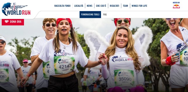 ci sarà la Cure Girl Sabrina a rappresentarci, a differenza delle precedenti edizioni, stavolta in Italia per tutti noi sarà possibile partecipare alla corsa solo tramite APP RUN, l’applicazione per smartphone appositamente realizzata per l’evento.
 This donation will go towards Spinal Research’s
 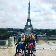 Assim como nós mudamos, mudam também as cidades, vinte e cinco anos depois eu voltava a terra da Rainha. Como estive aqui no começo da adolescência (e ainda andava) era uma cidade diferente. Londres foi uma experiência incrível pela receptividade, acessibilidade e intensidade. Receptividade por que pude sentir desde a minha chegada como os laços se fortalecem mesmo à distância, Lolly, seus irmãos Tony e Gary, e sua mãe Maureen me receberam como parte da família que realmente somos. A acessibilidade tornou possível cumprir a agenda realmente intensa que tivemos entre pubs, Paris e turismo ocasional. Nossa primeiro compromisso foi conhecer o pessoal incrível do escritório da Spinal Research, e
 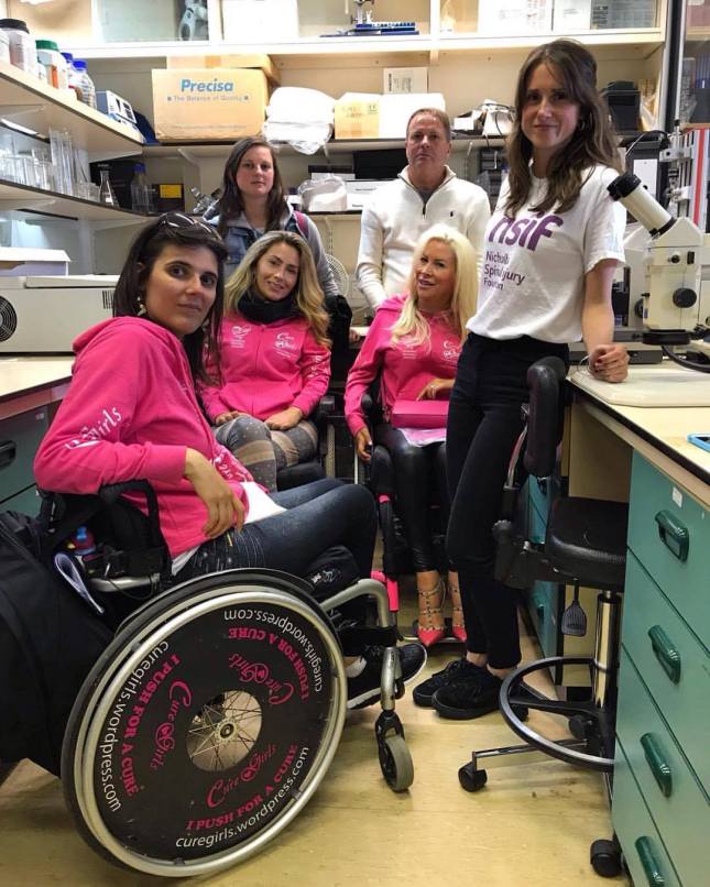 Aguardamos boas noticias para os próximos anos! Nesse dia conheci pessoalmente a Loredana, minha “cure girl sister” da Itália, que estava com uma equipe filmando o primeiro Doc das Cure Girls, espero que em breve todos possam sabem ainda mais sobre nossos projetos e anseios de 7 garotas ao redor do mundo lutando pela cura das lesões na Medula. Que desse encontro surja a possibilidade de tratamentos realmente eficazes para Lesões Medulares. Ainda sonho em assistir um show dos Rolling Stones no Hyde Park, para isso tanto Mick & cia quanto as Cure Girls precisarão da ciência caminhando ao nosso lado. “You can’t always get what you want, but if you try sometimes…”

 Stiamo aspettando con ansia buone notizie per gli anni a venire! Grazie a questo viaggio, ho incontrato Loredana, la mia “cure girl sister” dall’Italia. Speriamo che presto tutti conosceranno ancora di più i progetti e le speranze di sette ragazze provenienti da tutto il mondo, che combattono per la cura delle lesioni spinali. Ci auguriamo che da questo incontro si realizzi la possibilità di avere trattamenti veramente efficaci per le lesioni del midollo spinale.
 London was an incredible experience for its receptivity, accessibility and intensity. Receptivity because I could feel, since my arrival, how the bonds have grown stronger even at a distance — Lolly, her brothers Tony and Gary and their mother Maureen welcomed me as part of the family we really are. The accessibility made it possible for us to fulfill the really intense schedule we had between pubs, Paris and occasional tourism.
 of Dr. Liz Bradbury at Kings College, where she talked about her fifteen years of research and search for the healing of spinal cord injuries. We got to see incredible things which she uses in her studies. In the afternoon we went to the UCL lab which outlines another line of research, seeking to treat chronic injuries in humans and is funded by the Nicholls Spinal Injury Foundation. We spoke to Charlotte who is the Finance Manager of NSIF and also the researcher Professor Ying Li and the team.
  We’re looking forward for the good news in the years to come! On that day I personally met Loredana, my “cure girl sister” from Italy. Hopefully, soon everyone will know even more about the projects and hopes from seven girls around the world fighting for the healing of spinal cord injuries. May the possibility of truly effective treatments for the spinal cord injury arise from this meeting. I’m still dreaming of watching a Rolling Stones concert in Hyde Park — and for this to happen, both Mick & Co. and Cure Girls will need science walking by our sides. “You can’t always get what you want, but if you try sometimes…”

 Lo studio di Emily Burnside “Regulateable Chondroitinase ABC” gene therapy as a treatment for spinal cord injury” (Terapia genetica per la Regolabilità del Chase come trattamento delle lesioni midollari) potrebbe accelerare i tempi per la sperimentazione sull’uomo del Chase. La dr.ssa Burnside ha spiegato che uno dei problemi del Chase è che una volta somministrato rimane attivo per pochi giorni, mentre servirebbe che rimanesse attivo più a lungo, quindi andrebbe iniettato ripetutamente nel midollo. Nel suo studio hanno somministrato il Chase al midollo spinale lesionato di animali attraverso una strategia per modificare geneticamente le cellule nella zona della lesione in modo che le cellule stesse producessero il Chase. In questo modo si è risolto il problema di doverlo iniettare ripetutamente e si è ottenuto sia un forte contenimento del danno che un recupero funzionale importate sia in lesioni midollari a livello toracico che cervicale.
 ricercatore esperto che come il prof. Silver non ha mai perso la speranza nel cercare il modo di rigenerare il midollo spinale. Il dr. Lu è personalmente motivato a trovare una cura per le lesioni midollari croniche in quanto egli stesso è paralizzato dal 1996 a seguito di un incidente stradale. Egli ha condotto diversi studi molto importanti basati sull’uso di cellule staminali. In particolare in uno
 In the last few weeks the Cure Girls Loredana and Marina have been very busy with two annual events to raise money for the association

