San Diego, California – The annual meeting of the Society for Neuroscience (SFN) ran for six days here in November, as more than 30,000 researchers and academics from 90 countries presented over 15,000 science reports covering a huge variety of brain and spinal cord topics.
The meeting fills a giant convention center, row after row of bulletin boards displaying 3-ft. By 5 ft. data summaries of recent experiments; these are called posters, which are organized by theme. Each poster is displayed only for half a day; the main author is usually there to answer any questions from his or her peers. The cool thing about posters is that the work has not always been published in the medical literature, therefore giving the field a peek at what’s to come.
The rest of the convention floor includes hundreds of commercial vendors selling everything from mutated mice to multi-million dollar microscopes. One is struck by the enormous diversity of the neuroscience field, both in terms of the science itself, and of the international industry that sustains all of it.
There are of course many clinical or disease specific research areas, including studies of Alzheimer’s, stroke, pain, MS and visual degeneration. This year there were an abundance of discussions and posters on mosquitos (zika virus), football (concussions) and adolescents (autism).
I went on the lookout for clinical angles related to chronic spinal cord injury (SCI). Most of what is presented at SNF is not directly applicable to human disease or trauma. The agenda is driven by basic science, a myriad of hypotheses being tested in hopes of figuring out the central nervous system. It’s a biologist’s pleasure dome with a wide focus: gene editing, nervous system mapping, neural development, sensory and motor systems, cognition, neuroethics, addiction and plenty more. The meeting can be overwhelming, but navigation toward the areas of one’s interest has been made easier now with phone apps and online tools. If you’re inclined, have a look at this year’s program; you can search for a topic of specific scientist. SFN staff curated several schedules, including one for brain and SCI.
In this article we’re going to look at a few SNF science presentations I came across that have potential for chronic paralysis: 1) Modification of spinal cord scarring to allow nerves to grow across a non-permissive environment; 2) use of cell therapies in restoring function after SCI.
The scar:
After injury to the spinal cord, the damaged area loses a lot of nerve cells, which are cleared out by the immune response; a cavity forms and is eventually lined by a type of scar. Nerves have some capacity to grow after injury but this scar is a barrier. Jerry Silver, a scientist from Case Western in Cleveland, Ohio was the first to characterize the scar (chondroitin sulfate proteoglycans) and to imagine ways of getting rid of it. He and others found that by using a bacterial enzyme called chrondroitinase, they could chemically digest the scar – even in long term injuries. If you follow SCI research you have heard of this stuff, nicknamed ‘chase;’ it has been used in experiments to allow nerve axons to cross the scar and restore significant amounts of function. It’s a deceptively simple idea, just apply chase-juice to clear the path for regeneration. There are many issues with the juice, though, including how to deliver it safely in a human animal.
Previously, Silver used chase along with little nerve grafts to rewire and restore breathing function in tetraplegic lab animals. Said he in 2011, “Our work is to-date one of the most convincing demonstrations of the return of robust function after paralysis.”
I ran into Silver at an SCI-related poster session. He remains one of the most hopeful researchers when it comes to chronic SCI, and he was bubbling with enthusiasm for the “shocking recovery” seen in his most recent work: animals with what he called “super chronic” paralysis, one and a half years post injury, respiratory function was restored to “essentially normal” after getting chase and serotonin, a chemical needed for nerve transmission.
“This is the culmination of 30 years of work,” Silver said. “Apparently the longer we wait the better. I had some animals which we basically forgot about in the basement. We thought, why not try our treatment. It was astounding. Within two weeks there was complete recovery. Sometimes accidents can be good!”
Silver said he next wants to target systems other than respiration, such as hand function, or bladder, using chase or a peptide his lab has developed that prevents the growing tips of axons from getting stuck on sugary proteins of the scar.
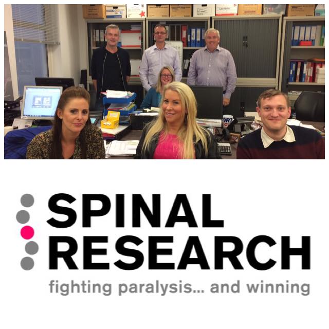
 When I ran into Silver he was observing a poster being presented by Emily Burnside, a member of the Elizabeth Bradbury lab at King’s College, London. Bradbury and her group are leaders in applying chase to SCI; she is co-principal investigator for major push, called CHASE-IT, to bring this stuff to clinic. The funding for this comes from the UK based Spinal Research charity.
Burnside’s poster, “Regulateable Chondroitinase ABC [aka chase] gene therapy as a treatment for spinal cord injury,” could hasten time to the clinic. She explained that the lab had previously delivered chase to the injured spinal cord of animals using a gene modification strategy by way of a virus that ferried the gene code for chase to neurons in the injury site; chase is then produced by the nerve cells themselves. One of the issues with chase is that it doesn’t last long once administered, so it has to be given repeatedly. Gene therapy addresses that. “This treatment [gene therapy vector] resulted in dramatic reduction in pathology and significant improvements in functional recovery following clinically relevant spinal contusion injury at both thoracic and cervical levels in adult rats,” the poster noted.
A potential problem with a viral delivery system is that once the cells are turned on to make chase, they can’t be shut off. Too much chase may produce unwanted effects. So Burnside used a second vector to introduce a sort of on-off switch. “This gives us more control over chase, and allows us to optimize its timing,” said Burnside. “It is a step toward clinical relevance of the enzyme.”
Bradbury and her team were involved in several other posters. One presented data on a primate SCI model, using chase plus Schwann cell transplants; the treated animals improved almost to normal. This project is led by James Guest at the Miami Project to Cure Paralysis; Guest is principal investigator for a human trial in Miami of Schwann cell transplants in patients at least one year post injury.
I came across another poster on scar, this one from the UCLA lab of Michael Sofroniew, who has been saying for years that it’s wrong to blame the scar for the mediocre regenerative effort of spinal cord axons. Here, he and his lab mates show more data that the scar is not the bad guy, in fact, it actually helps regeneration. They used a bunch of growth additives to get axons to grow through the toxic scar area, but they did not do as well if the scar itself was removed. From the poster detail: “… preventing astrocyte scar formation, attenuating scar-forming astrocytes, or deleting chronic astrocyte scars all failed to result in spontaneous regrowth of transected corticospinal, sensory or serotonergic axons through severe spinal cord injury lesions. In striking contrast, sustained local delivery … of required axon-specific growth factors not present in SCI lesions, plus growth-activating priming injuries, stimulated robust … sensory axon regrowth past scar-forming astrocytes and inhibitory molecules in SCI lesions. Preventing astrocyte scar formation worsened this stimulated axon regrowth … Our findings show that contrary to prevailing dogma, astrocyte scar formation aids rather than prevents CNS axon regeneration.”
Cells therapies:
Paul Lu is a researcher in the San Diego lab of Mark Tuszynski, a veteran investigator who, like Silver, has never lost hope in the concept of spinal cord regeneration. Lu is motivated by personal reasons. He was paralyzed below the waist in an auto accident in 1996 while in grad school. He changed his major from botany to neuroscience, joined the Tuszynski group and has been responsible for some eye-popping stem cell research in an SCI animal model. A 2012 study showed that after implantation of neural stem cells, along with a cocktail of growth-promoting chemicals, spinal cord nerve fibers grew abundantly, and at great distances from the injury site. Lu saw no meaningful recovery but hopes that’s being worked out.
The lab reported at SFN that they had transplanted human neural stem cells into a primate model, a major step toward clinical usefulness. Adult rhesus macaques underwent C7 lateral hemicontusions [most common type of injury, but only one side affected] or lateral hemisection lesions [cut, not bruised]. The human stem cells were grafted into the SCI sites between 2 and 12 weeks after injury. Each animal got 20 million cells, suspended in a fibrin matrix and growth factor cocktail. Surviving grafts differentiated into both neurons and glia; hundreds of thousands of new axons grew, some growing as far as two inches. The study notes that the delivery of cells must be optimized before this can be tried in humans.
The Tuszynski lab, though without Lu, presented a poster at SNF showing that multipotent neural progenitor cells (NPC) supported axonal outgrowth and improved functional outcomes in a cervical contusive injury model. That was a sub-acute experiment, with cells transplanted at two weeks. The lab stated that they are now assessing NPC grafts in models of chronic contusive injury.
Another cool area Lu and the Tuszynski group are working on is using light sensitive cells (optogenetics) to a) make better connections; and b) to allow them to turn cell functions on and off during experiments. The lab is also on the trail of “master regulators,” the gene codes that could activate programs for axon regeneration.
At SFN, Lu told me the next big improvement in regeneration of stem cells will be the cells. He’s already experimented with induced pluripotent stem cells (iPSC), which are cells from an animal’s own body that are put in reverse, essentially becoming a type of stem cell that can take on any cell form. “New tools allow us now to make phenotype specific neural cells,” said Lu, which means he can make a cell that has the most desirable features.
There were many posters about iPSC. While the cells may have a high safety profile because they come from a patient’s own body, which also makes null the ethical issues regarding embryonic or fetal stem cells, iPSC carry some of the same baggage as other stem cells: they can form tumors. A group from Japan, led by Masaya Nakamura from the Keio University School of Medicine in Tokyo, is already to using human iPSC/oligodendrocyte precursors in animal models to show that the cells promote remyelination and that iPSC grafts integrate with host neuronal circuits and form synapses. On a poster here, the group showed that two lines of iPSC cells promote motor recovery but one forms tumors. A goal of the work, besides repairing SCI damage, is safety, that is, to develop genetic quality controls to make sure a particular iPSC line does not overproduce itself and make tumors.
The Michael Fehlings lab from the University of Toronto is actively looking at many types of interventions for SCI, including iPSC. In a poster at SFN, his group transplanted pluripotent stem cell derived neural precursor cells that were further modified to secrete a potent growth promoting molecule called GDNF. Animals got the cells two weeks after injury, so this is not considered an approach to chronic SCI, but the GDNF animals showed more recovery than those treated with precursor cells only.
Maybe there won’t be a need to add cells from the outside. Researchers are now finding ways to manipulate cells already in the body — to expand them, and perhaps to reprogram them take on new functions.
Lu noted the work of Chun-Li Zhang, at UT Southwestern, who has reprogrammed astrocytes in spinal cord scar cells to neurons. Zhang presented at an SFN symposium on creating spinal motor neurons from reprogrammed adult human fibroblasts (skin cells); this has a more obvious application in ALS but could have a role to play in spinal cord injury. Zhang also showed data on reprogramming neural cells in vivo – in a living animal – with potential in a spinal cord injury model. From the abstract: “Our ability to successfully produce a large population of long-lived and diverse subtypes of new neurons in the adult spinal cord provides a cellular basis for regeneration-based therapy for SCI.”
by Sam Maddox

 Questa donazione andrà al progetto CHASE-IT di Spinal Research per sviluppare una terapia efficace per la lesione del midollo spinale utilizzando le proprietà di potenziamento della neuroplasticità dell’enzima batterico condroitinasi.


 Few weeks ago the film “Me before you” has been released in England; it is based on the book by the English writer Jojo Moyes which I finished reading some days ago. This is about Will Traynor, a handsome, rich, active young man, with a great career, who finds himself quadriplegic following a spinal cord injury (quadriplegia = paralysis in all four limbs).
 “I don’t want you to be tied to me, to my hospital appointments, to the restrictions on my life. I don’t want you to miss out on all the things someone else could give you. And, selfishly, I don’t want you to look at me one day and feel even the tiniest bit of regret or pity and…”


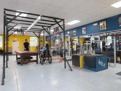 y. Using external stimulation for the nervous system to promote reorganization they are reminding the nerves and muscles how to work again. Muscle spasms are used to build muscle mass and control, using the spasms rather than having to fight against them. They do a lot of weight bearing activities; this in itself promotes healthy bones and fitness. They do not say people will be hopping and skipping out of the door. They do say the best case is a client can regain function and continue to improve as the exercises help a client’s body to remember how to move. Worst case is the client will just leave more independent and healthy. The health benefits are great and this is something that is very much needed for a SCI person. Taken from Project walks site the below are results.
 e this kind of therapy benefits all of us. It’s not a cure for paralysis but is a great way to get our bodies fit, possibly improving movement and sensation and all of the above.As a Cure Girl I believe this is something that should be set up around the world, we all could use a Project Walk. I would like to thank Liza, Amanda and Brock for showing my family and I around and I will definitely be back.